

Ask an Expert Q&A: Vivek Gopalakrishnan
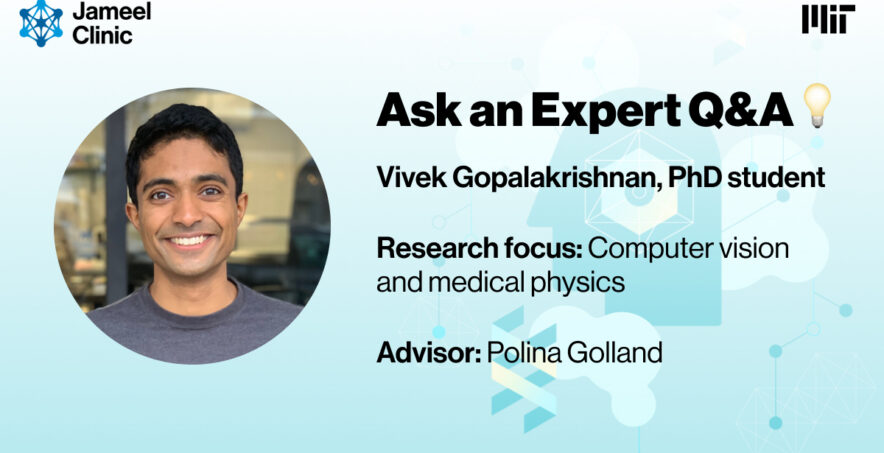
Vivek Gopalakrishnan is a fifth year PhD student in the Harvard-MIT Program in Health Sciences and Technology and a ‘25 MIT HEALS Fellow, advised by Jameel Clinic PI Polina Golland. His research combines computer vision and medical physics to extract quantitative 3D/4D information from routine 2D medical images (X-ray and ultrasound), enabling new directions in image-guided interventions and surgical robotics.
1) What’s the biggest question you are trying to answer in your work?
Humans possess the remarkable ability to perceive 3D structure and motion from 2D images of the natural world, but this doesn’t extend innately to image-guided interventions, where clinicians are forced to navigate using grainy X-rays that carry no depth information. Can we instead build machines that can do this reconstruction to within a millimeter of accuracy, in real time, without failing on a single patient?
2) What’s something in your research that’s been exciting or surprising lately?
A surprising direction is that overfitting, normally the cardinal sin of machine learning, is exactly the right move here: rather than fitting a training set of arbitrary humans, we deliberately overfit a model to the single patient undergoing the procedure. Nearly every surgical patient already has a preoperative CT or MRI, so we render synthetic X-rays from their own anatomy and train this model in about five minutes, with no change to the current clinical workflow.
3) What is the biggest challenge in your area of research?
Biomedical imaging datasets are relatively small, often containing only tens to hundreds of patients, so we cannot scale our way to generalizability the way the rest of machine learning does. Building data engines that produce synthetic data through physics-based simulation has become indispensable in my research, because if we can’t simulate it, we can’t train on it.
4) If your research succeeds, how could it help patients or medicine?
Minimally invasive procedures currently demand clinicians who have trained for decades to reconstruct 3D anatomy in their heads. Augmented vision systems would let more junior clinicians achieve similar proficiency, and eventually lead to surgical robotic systems that can navigate autonomously, which would ultimately bring these interventions to patients who have no access to them today.
5) What papers or whose work have you been reading lately that you’d like to give a shoutout to?
Perhaps unexpectedly, some of the research most relevant to me comes from computer graphics. In particular, I’ve been enjoying Marilyn Keller’s research on biomechanically accurate 3D humans (https://hit.is.tue.mpg.de/), which augments the statistical body models used for character animation with anatomically realistic skeletons and soft tissue. Being able to reposition a patient’s volumetric scan into a new, anatomically valid pose could be very useful for building machine learning models that are robust to patients in non-standard orientations (e.g., after trauma).
—
Ask & Expert Q&A is a monthly Q&A series featuring experts doing cutting-edge work at the intersection of artificial intelligence and health who are affiliated with the MIT Jameel Clinic. If you have any questions for our researchers or are interested in being featured, please reach out to jclinic-info(at)mit.edu.
